
Arthritis, a common condition affecting millions worldwide, doesn't just manifest as physical discomfort. An accurate diagnosis often requires imaging that unveils what the eye cannot see. Among the various tools available, Magnetic Resonance Imaging (MRI) stands out for its precision and depth.
Unlike conventional X-rays, MRIs can provide a comprehensive view of the joint and surrounding tissues. This ability to look deeper means that not only can we spot arthritis, but we can also understand its severity and impact on the body.
- Introduction to MRI and Arthritis
- How MRI Works in Detecting Arthritis
- Types of Arthritis Visible on MRI
- Benefits of MRI in Diagnosing Arthritis
- Challenges and Limitations of MRI
- Practical Tips for Patients and Clinicians
Introduction to MRI and Arthritis
Magnetic Resonance Imaging, better known as MRI, has transformed the healthcare landscape since its inception. Offering a detailed and non-invasive view of the body's internal structures, it allows medical professionals to see beyond the surface. When it comes to identifying and understanding arthritis, MRIs become an integral part of the diagnostic toolkit. They can capture images of both the bones and soft tissues such as cartilage, ligaments, and the surrounding joint fluid. This ability is vital for assessing conditions like arthritis, which often affects these specific areas.
Unlike traditional X-rays that primarily assess bone condition, MRIs can reveal subtle changes and early signs of joint damage. This is especially crucial in diseases such as rheumatoid arthritis and osteoarthritis. An MRI can detect synovitis, bone marrow lesions, and cartilage degradation long before they become visible on an X-ray. For patients experiencing symptoms uncharacteristic or unexplained by other diagnostic tools, MRI can be the clarion that calls for a deeper examination.
Recognizing Different Types of Arthritis
The power of MRI lies in its versatility. It doesn't just point out the presence of arthritis; it differentiates between types. Each arthritis type presents a unique set of challenges and symptoms, and knowing which kind you’re dealing with helps tailor the treatment plan. Osteoarthritis, often considered 'wear-and-tear' arthritis, showcases cartilage thinning and changes in bone density – all discernible on MRI. Conversely, rheumatoid arthritis displays inflammation and potential joint erosion, which is crucial for early interventions. For clinicians, these detailed images guide the severity assessment and monitoring disease progression over time.
"MRIs have revolutionized the way we understand arthritis. What was once a black-box diagnosis now has a window into its workings." —Dr. Elizabeth Dunne, Radiologist at Johns Hopkins Hospital
While patients and doctors marvel at the insight MRIs provide in diagnosing arthritis, it is equally important to assess their practicality. They are entirely non-invasive, involving no radiation, unlike CT scans. This suitability for repetitive use makes MRIs ideal for monitoring conditions and adjusting treatment plans. Patients facing chronic joint health issues benefit tremendously from this innovation, allowing them to track progression and effectiveness of their therapy plans.
How MRI Works in Detecting Arthritis
When exploring the inner workings of Magnetic Resonance Imaging, or MRI, one taps into a blend of sophisticated technology and biology. MRI leverages powerful magnets and radio waves to generate detailed images of the body's interior, diving beneath layers that remain elusive to traditional X-rays. This imaging prowess proves crucial in detecting arthritis, providing a visual narrative of joint health that is remarkably rich in detail. The ability to visualize both soft and hard tissues helps radiologists spot even the subtle hints of inflammation or degeneration typical of arthritic changes.
MRIs excel in their capacity to highlight differences in tissues due to the strong magnetic fields that cause nuclei in cells to resonate at specific frequencies. These resonances vary based on the tissue's physical makeup, so when these signals are processed, an image reflecting the various textures and structures within a joint emerges. This sensitivity is particularly effective in distinguishing between healthy cartilage and areas where arthritis may be taking its toll. Interestingly, MRI can detect not only the traditional markers of arthritis, like bone erosion, but also early signs such as cartilage thinning or soft tissue changes. It's this precision that elevates its role in diagnostics.
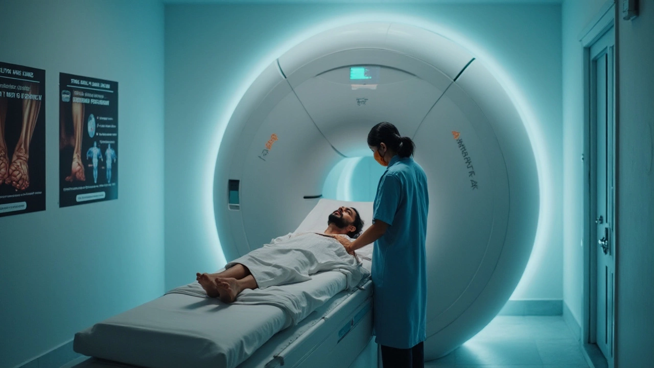
The process begins with placing the patient in the cylindrical MRI machine, where they must remain still—a challenge for those in discomfort yet necessary for clear imaging. The machine's magnets align hydrogen atoms in the body before radiofrequency pulses knock them out of place. As they realign, they emit energy, captured and converted into images by the MRI scanner. This technique covers joints in three dimensions, which allows doctors to interpret intricate details from different angles, potentially transforming how orthopedic specialists formulate treatment plans.
According to Dr. James Andrews, a noted orthopedic surgeon, "MRI findings can significantly influence treatment decisions, allowing for customized approaches tailored to the underlying causes of joint pain."
Understanding the types of arthritis that MRI can visualize is key. The machine has proven especially effective with inflammatory arthritis such as rheumatoid arthritis, highlighting synovial inflammation before full-blown joint damage occurs. Similarly, with osteoarthritis, while it primarily affects cartilage, MRI's ability to visualize bone and soft tissues offers an encompassing perspective, identifying underlying conditions other methods might miss.
While the advantages are clear, MRIs are not without their limitations. They can be expensive and sometimes inaccessible to everyone. But for many, their diagnostic effectiveness offsets these words of caution, providing insights that could prevent further degeneration through early intervention. Those considering an MRI should consult with their healthcare provider about the potential benefits specific to their situation.
It's this combination of advanced technology and biological insights that underscore the role MRI plays in detecting arthritis. As the tools and techniques evolve, the clarity with which we can diagnose conditions like arthritis will only improve, giving patients a clearer path to effective treatment and potentially better outcomes.
Types of Arthritis Visible on MRI
When it comes to diagnosing arthritis, identifying the specific type or variant is crucial for effective treatment. MRIs have revolutionized how we view and understand the internal elements of our bodies, enabling clinicians to differentiate between various forms of arthritis with greater precision. This imaging method is particularly adept at highlighting subtle differences in tissue composition, inflammation levels, and cartilage health that characterize distinct types of arthritis.
Osteoarthritis, the most prevalent type, often shows up on an MRI as a loss of joint space, cartilage wear, and possible bone spurs. Unlike the standard X-ray, MRIs capture these changes in greater detail, making it possible to identify even the early onset before significant pain develops. This clarity helps tailor prevention strategies, slowing progression through lifestyle adjustments and medication early on.
Rheumatoid arthritis, an autoimmune condition, presents quite differently under MRI scrutiny. This type of arthritis is driven by inflammation, and MRIs can highlight synovial membrane thickening, which is a classic sign. Joint damage, swelling, and sometimes even signs of early bone erosion can also become evident. Spotting these markers early is instrumental in managing the disease effectively. Dr. Susan Smith, a renowned rheumatologist, once noted,
"MRI allows us to see under the skin, into the inflammatory process, making it a key tool for establishing early intervention paths."
Less common yet significant are conditions like psoriatic arthritis and ankylosing spondylitis. With psoriatic arthritis, an MRI might reveal transformative changes where tendons attach to bone, known as enthesis, a hallmark feature of the disease. Ankylosing spondylitis can be spotted through detected inflammation in different areas like the lower back and pelvis. MRIs can even uncover the development of new bone structures, showing the body’s misguided attempts to fuse the spine.
Statistically, patients seeking orthopedic consultation have shown that around 70% saw clearer pathology after an MRI scan. This revelation not only aids correct diagnosis but also instills confidence in treatment plans devised based on such detailed images. Many patients report feeling empowered when they visually connect to their unexplained pain on scan reports.
Benefits of MRI in Diagnosing Arthritis
Magnetic Resonance Imaging, or MRI, revolutionizes the way we understand and diagnose arthritis. It offers a glimpse into the intricate world of our joints, free of invasive procedures. One of the most significant benefits is its ability to capture soft tissues in remarkable detail. This subtlety allows clinicians to assess inflammation, pinpoint structural changes, and observe cartilage damage, aspects often missed by traditional X-rays. By diving into the details, MRI provides a comprehensive view, thus ensuring each assessment is both detailed and profound. This is particularly crucial for early-stage arthritis, where signs are subtle.
Another advantage of MRI is its unparalleled safety profile. Unlike some imaging methods, MRI does not involve exposure to ionizing radiation. This makes it a safer alternative, especially for children or individuals requiring multiple scans. The technology relies on powerful magnets and radio waves, harnessing them into images. This radiation-free approach overcomes many concerns traditionally associated with imaging methods. It's an especially important consideration for patients already managing chronic conditions or those with a history of cancer.
In particular cases, the detailed imagery provided can influence and even change clinical management. Clinicians are equipped to better plan a course of action—be it medicinal intervention, lifestyle changes, or surgical procedures. An accurate diagnosis dictates an accurate treatment plan, which often results in better outcomes. This tailored approach extends to tracking the disease's progression over time. Regular MRIs offer a documented timeline of how arthritis evolves in response to treatments. This level of detail can empower patients, knowing that their treatment is not just reactive but proactive.
For those involved in orthopedic research, MRI serves as an invaluable resource. It allows the scientific community to verify findings and push boundaries in understanding joint conditions. In an enlightening statement, Dr. Sheila Brandford of the Joint Imaging Institute noted,
"MRI is not only a diagnostic tool but a window into joint dynamics. It changes how we envision arthritis therapy and challenges us to think beyond the typical."Her thoughts reflect the broader sentiment within the scientific community. The reliability of MRI findings is continually pushing boundaries in how diseases are understood and managed globally.
While MRIs aren't always the first line of defense, their unique advantages make them indispensable in specific scenarios. Whether it's obtaining a baseline view, delving deeper into disease mechanics, or ensuring safety without the burden of radiation, their benefits are clear. Patients and clinicians both stand to gain immense value from utilizing this technology. By incorporating MRIs into the diagnostic process, they not only enhance diagnosis accuracy but drive successful treatments. This marks a significant advancement in personalizing healthcare and improving patient outcomes.

Challenges and Limitations of MRI
While Magnetic Resonance Imaging (MRI) is an excellent tool for diagnosing arthritis, it is not without its challenges and limitations. One of the most significant considerations is the cost. MRIs can be quite expensive, making them less accessible for individuals without adequate insurance coverage. This financial barrier can hinder timely diagnosis and treatment. It's also worth mentioning that while an MRI provides detailed images, interpretation can be complex, requiring specialized training and experience to accurately understand what the images reveal about joint health.
Another concern revolves around the time it takes to conduct an MRI scan. Patients need to remain still for extended periods, which might be uncomfortable, especially for those already experiencing joint pain. Claustrophobia is also a known issue, as the MRI machine can be quite confining. Alternatives like open MRIs exist, but they may not be available everywhere. Despite its effectiveness, an MRI might sometimes miss subtle changes in bone structure, which are better captured by X-rays. Thus, while it provides a good look at tissues, it is not always the definitive diagnostic tool for certain types of arthritis.
According to Dr. Lisa Thompson, a leading orthopedic specialist, "MRIs provide a wealth of information, but not all conditions require such detailed imaging. It's crucial to weigh the need against practical issues like cost and patient comfort."
Technical limitations of MRI machines can also lead to diagnostic issues. They may produce artifacts—distortion in images—that complicate the diagnosis. This is especially prevalent in patients with metal implants, as MRIs are sensitive to metal and can sometimes provide inaccurate readings. Additionally, MRI scans are not suitable for everyone; patients with pacemakers or certain types of metal implants might be advised against them due to potential risks.
Another limitation includes the specificity of orthopedic diagnosis through MRI. Although they can highlight inflammation and soft tissue pathology, MRI scans might not effectively distinguish between different types of arthritis without a comprehensive clinical correlation. For instance, while inflammation in the joint area can indicate multiple types of arthritis, it does not specify which one without additional context. Clinicians must consider the patient's symptoms and medical history alongside MRI findings to reach a conclusive diagnosis.
Practical Tips for Patients and Clinicians
For patients experiencing joint discomfort, understanding the role of MRI in diagnosing arthritis is crucial. Before undergoing the procedure, it's essential to communicate openly with your healthcare provider about any metallic implants or claustrophobia concerns due to the MRI's enclosed nature. A comprehensive discussion will help in preparing both mentally and physically for the scan. It's also beneficial to inquire about how the scan results will be interpreted in correlation with your symptoms. Knowing what to expect in terms of sensations and noises during the procedure can help ease the anxiety that often accompanies medical imaging. Similarly, being informed about the potential outcomes and next steps following the MRI can give patients the confidence to engage more proactively in their treatment plans.
Clinicians, on the other hand, should strive to ensure that their patients feel supported and informed throughout the diagnostic process. Clear communication about the benefits and limitations of using MRI as a diagnostic tool for arthritis is essential. Clinicians should emphasize that while MRIs are highly effective in highlighting inflammation and other tissue changes, the results need to be considered as part of a broader diagnostic picture, including clinical examination and patient's medical history. Regular updates on technological advancements in orthopedic diagnosis can also enrich a clinician's capacity to provide cutting-edge care.
"MRI is one of the most powerful diagnostic tools available in orthopedics today, allowing us to examine soft tissues with extraordinary detail," says Dr. Erica Carter, a renowned orthopedic specialist.
Patients and clinicians might also find it useful to engage with MRI results using a step-by-step approach. Start by identifying the most affected joints and explore any visible abnormalities. Recognizing the specific type of arthritis visible on the scan can guide treatment decisions. Encouraging patients to share any changes in symptoms, especially after an MRI, can enhance the dialogue between patient and provider, facilitating a more tailored treatment approach. Clinicians should remain open to new technologies and methods in imaging that may offer even greater insights into patient health and treatment efficacy. Patients, meanwhile, benefit from staying informed and proactive, ensuring they understand each phase of their health journey.
Steps for Better MRI Experience
Whether you're a first-time MRI recipient or a healthcare provider, these practical steps can greatly enhance the overall experience and outcomes:
- Ensure detailed patient history is shared with the imaging technician to tailor the scan parameters accordingly.
- Clinch the importance of remaining still during the scan to avoid image distortion, which can significantly impact diagnostic accuracy.
- Utilize relaxation techniques such as deep breathing or meditation to ease stress prior to the procedure.
- Encourage a post-scan consultation where patients can discuss immediate impressions of their MRI results with their doctors, paving the way for a clearer understanding and less miscommunication.
The introduction of newer MRI technologies continues to improve the clarity and accuracy of diagnosing conditions like arthritis. In the coming years, expect even greater integration of MRI with advanced analytical tools to not only detect but predict arthritis progression, thus opening avenues for earlier interventions and optimized patient outcomes.